
The moment a physician suspects cancer, every diagnostic decision carries weight. While biopsies provide cellular confirmation, magnetic resonance imaging offers something equally critical: spatial understanding. It reveals tumor size, location, invasion patterns, and relationships to surrounding structures-information that directly shapes whether a patient undergoes surgery, radiation, chemotherapy, or a combination of approaches.
For patients navigating a cancer diagnosis, understanding when and why MRI becomes necessary can reduce anxiety and help them participate more actively in treatment decisions. The technology has evolved significantly, offering clearer images with shorter scan times, but knowing what to expect and how results influence care remains essential knowledge.
How MRI Differs from Other Cancer Imaging
Computed tomography scans excel at detecting abnormalities quickly, particularly in the chest and abdomen. PET scans highlight metabolically active areas, making them valuable for staging. MRI, however, provides superior soft tissue contrast, making it the preferred choice for brain tumors, spinal cord involvement, pelvic cancers, and musculoskeletal malignancies.
The distinction matters because treatment planning requires precise boundaries. A brain tumor that appears to be 2 centimeters on CT might reveal microscopic extension into eloquent cortex on MRI, fundamentally altering surgical approach. Similarly, rectal cancer staging depends on MRI’s ability to show whether the tumor penetrates through the bowel wall or invades adjacent organs-information that determines whether a patient needs preoperative chemotherapy or can proceed directly to surgery.
Contrast Enhancement and Tumor Behavior
Most oncological MRI protocols include contrast agents, typically gadolinium-based compounds injected intravenously. Tumors develop abnormal blood vessel networks to support rapid growth, and these vessels allow contrast to accumulate in patterns that help radiologists distinguish benign from malignant tissue, assess tumor aggressiveness, and monitor treatment response.
Patients with kidney impairment require careful evaluation before contrast administration, as reduced renal clearance increases risk of complications. Alternative protocols exist, though they may provide less diagnostic information. This trade-off represents one of many clinical decisions where individual patient factors shape imaging strategy.
Specific Cancers Where MRI Proves Essential
Breast cancer staging increasingly incorporates MRI, particularly for young women with dense breast tissue where mammography has limitations. The technique identifies multifocal disease-additional tumor sites within the same breast-in approximately 15 to 20 percent of patients, information that can shift treatment from lumpectomy to mastectomy.
Prostate cancer presents another area where MRI has transformed practice. Multiparametric MRI combines multiple imaging sequences to create a detailed prostate map, allowing targeted biopsies of suspicious regions rather than random sampling. This approach increases detection of clinically significant cancers while reducing diagnosis of indolent tumors that may never require treatment.
Liver metastases, common in colorectal and neuroendocrine cancers, often appear more clearly on MRI than on CT. For patients considering surgical resection of liver tumors, MRI helps surgeons plan which segments to remove and assess whether sufficient healthy liver will remain. In healthcare systems offering advanced integrative approaches to cancer treatment, imaging results inform not just surgical decisions but also nutritional support strategies and rehabilitation planning.
Accessing MRI Services in Regional Healthcare Systems
The United Arab Emirates has invested substantially in medical infrastructure, creating regional centers that combine diagnostic imaging with comprehensive cancer care. Facilities like Cleveland Clinic Abu Dhabi integrate imaging services directly with oncology departments, allowing rapid interpretation and treatment planning. For patients researching options, understanding both the cost of mri in abu dhabi and the availability of subspecialized radiologists who focus specifically on oncological imaging helps in selecting appropriate care pathways.
The broader development of specialty healthcare in the region reflects recognition that cancer diagnosis requires more than equipment-it demands expertise in protocol selection, image interpretation, and communication with treating physicians. Modern cancer centers coordinate multidisciplinary conferences where radiologists present imaging findings alongside pathologists, surgeons, and medical oncologists, ensuring that every perspective informs the treatment recommendation.
What Patients Experience During Oncological MRI
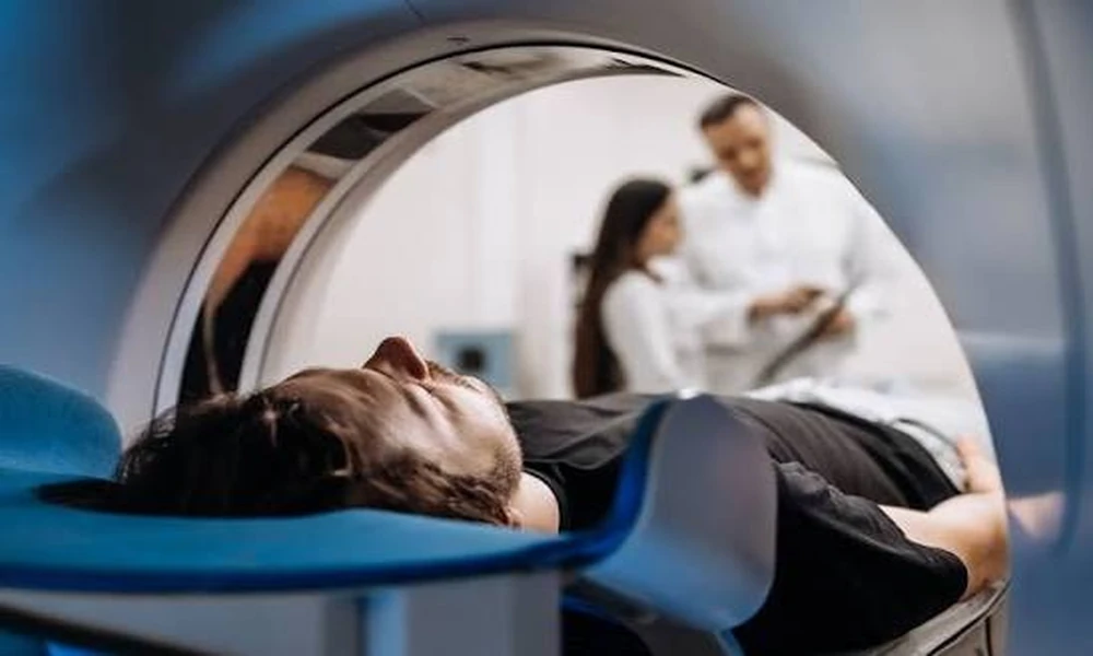
Scan duration varies by body region and protocol complexity, typically ranging from 30 to 90 minutes. Patients lie motionless inside a cylindrical magnet while radiofrequency pulses generate images. The machine produces loud knocking sounds despite hearing protection, and the enclosed space challenges those with claustrophobia.
For cancer patients already managing treatment side effects, the physical demands of remaining still can prove difficult. Technologists can provide positioning aids, breaks between sequences, and clear time estimates. Open MRI systems offer more space but may sacrifice some image quality, presenting another clinical trade-off that depends on the specific diagnostic question and patient tolerance.
Repeat Imaging and Radiation Avoidance
Unlike CT scans, MRI uses no ionizing radiation, making it suitable for repeated monitoring. Cancer patients often require imaging every two to three months during active treatment to assess response, then at lengthening intervals during surveillance. The cumulative radiation exposure from multiple CT scans, while generally acceptable given the benefits, can be avoided when MRI provides equivalent information.
This advantage particularly benefits pediatric cancer patients and young adults who face decades of follow-up imaging. The investment of additional scan time becomes worthwhile when considering lifetime radiation exposure and long-term secondary cancer risk.
How Imaging Results Shape Treatment Sequencing
Neoadjuvant therapy-chemotherapy or radiation given before surgery-has become standard for many solid tumors based partly on imaging findings. MRI performed after several chemotherapy cycles shows whether tumors are shrinking, stable, or progressing. Dramatic size reduction might make previously inoperable tumors resectable. Lack of response prompts treatment changes before surgery, avoiding ineffective therapy and surgical delay.
Regional healthcare development, including specialized facilities designed to support complex oncological care, has made these sophisticated treatment sequences more accessible to patients who previously would have needed to travel internationally. The integration of imaging services with surgical and medical oncology creates workflows where results translate rapidly into action.
Functional MRI Techniques in Cancer Care
Beyond anatomical imaging, functional techniques assess tumor biology. Diffusion-weighted imaging measures how water molecules move through tissue, providing information about cellular density. Highly cellular tumors restrict water movement, appearing bright on specific MRI sequences. This technique helps distinguish tumor recurrence from post-treatment scarring, a common clinical dilemma.
Perfusion imaging evaluates blood flow through tumors, which correlates with aggressiveness and treatment response. Spectroscopy analyzes the chemical composition of tissue, detecting metabolic signatures associated with active cancer. While not yet standard everywhere, these techniques represent the direction of precision oncology, where treatment intensity matches biological behavior rather than relying solely on tumor size and stage.
Preparing for Results and Next Steps
Radiologists typically provide preliminary readings within 24 to 48 hours, with formal reports following. For cancer patients, waiting for results generates significant anxiety. Understanding that imaging provides one data point among many-pathology, molecular testing, overall health status-helps maintain perspective.
When results arrive, patients benefit from prepared questions: What exactly did the scan show? How does this compare to previous imaging? What are the next steps regardless of the findings? Does this change the treatment plan or timeline? Oncology teams expect these questions and should provide clear answers that acknowledge uncertainty where it exists rather than offering false reassurance.
The relationship between imaging technology and cancer outcomes continues evolving. Better images lead to more precise treatments, which improve survival and quality of life. For patients facing cancer diagnosis or monitoring, understanding MRI’s role transforms it from an intimidating procedure into a powerful tool that, while imperfect, provides essential information guiding every subsequent decision.